 Pori

- A seborrheic keratosis is a harmless, waxy or warty growth that looks stuck onto the skin, usually tan, brown or black, and common from middle age on.
- They are not contagious and are not considered precancerous; people simply get more of them with age, and they often run in families.
- Melanoma can closely mimic a seborrheic keratosis, so a spot that is new, changing, bleeding, very dark or different from your others needs a dermatologist.
- Removal is optional and done in a clinic by freezing, scraping or electrosurgery; the AAD says not to remove one yourself, and treated skin can lighten.
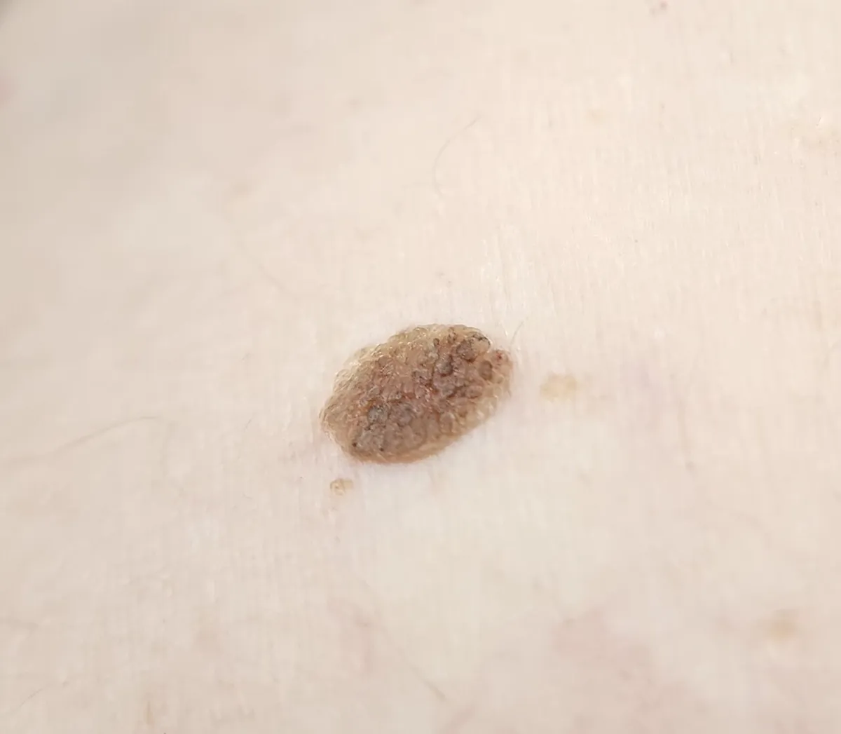
A seborrheic keratosis is a harmless skin growth that looks waxy, scaly or warty and seems pasted onto the skin, like a dab of brown candle wax or a barnacle. It is usually tan, brown or black, from a few millimeters to more than an inch across, and appears on the chest, back, face, scalp or neck from middle age on. Melanoma can look similar, so any new, changing, bleeding or unusual spot needs a doctor's eye.
Pori does not diagnose skin conditions or check spots. Its selfie scans and photo diary can help you keep dated, consistent photos to show a dermatologist and to see whether a spot or a treatment area is changing over time.
What does a seborrheic keratosis look like?
The American Academy of Dermatology (AAD) says these growths tend to start as small, rough bumps, then slowly thicken and develop a warty surface, and to have a waxy, stuck-on-the-skin look. That "stuck-on" quality is the classic clue: the spot looks as if you could lift it off with a fingernail. The AAD compares some to dabs of warm, brown candle wax and others to stucco splattered on the skin.
- Color: most are tan or brown, but the AAD notes they range in color from white to black. DermNet adds skin-colored, yellow, grey and mixed shades.
- Size: from a fraction of an inch to larger than a half-dollar. DermNet gives a range of about 1 mm to several centimeters.
- Surface: smooth and waxy, crumbly, or rough and warty.
- Feel: they don't hurt, though some itch, and they can catch on clothing, bra straps or jewelry.
- Where: the chest, back, stomach, scalp, face and neck. They don't grow on the palms or soles, and MedlinePlus adds the lips to that list.
How does it look on brown and black skin?
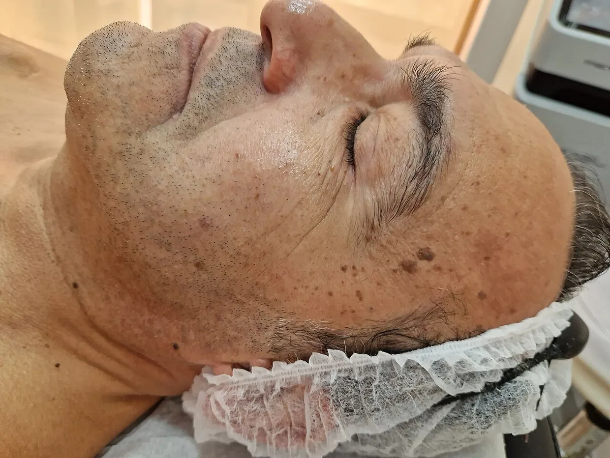
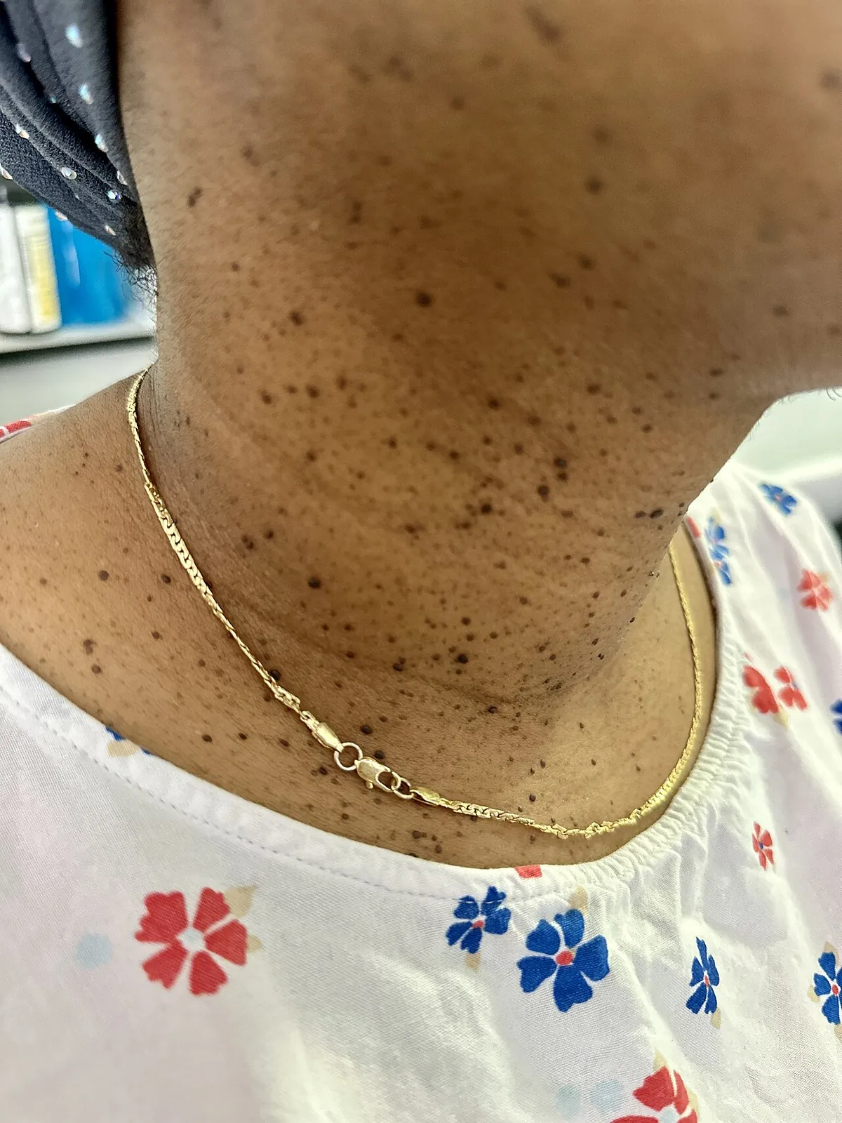
The AAD notes that these growths also develop in people with medium to dark skin, where they tend to be small and to appear around the eyes. Many small, dark, smooth bumps on the cheeks, around the eyes and on the neck are often dermatosis papulosa nigra, a variant DermNet describes as small, stalked, heavily pigmented lesions on the head and neck, mainly in people with darker skin. We cover it in its own guide to dermatosis papulosa nigra.
Who gets seborrheic keratoses, and are they contagious?
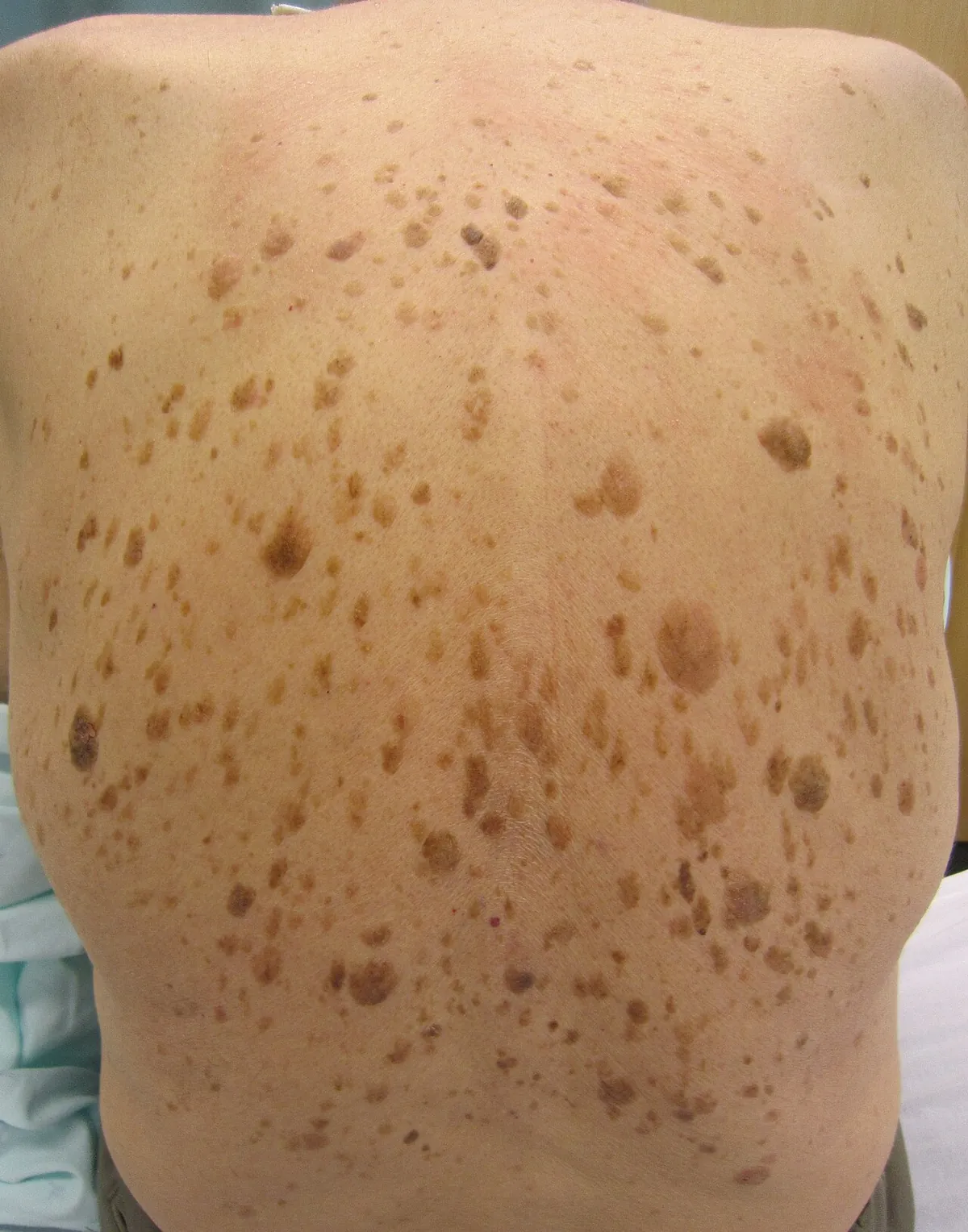
Almost everyone gets at least one with age. DermNet reports that over 90% of adults over the age of 60 years have one or more, and that they usually start appearing in the 30s or 40s. The AAD calls them the "barnacles of aging" and says children rarely have them.
The cause is unknown. According to the AAD, they seem to run in families, and some studies link them to sun exposure, though they also grow on skin that is always covered. Some people develop them during pregnancy or after estrogen replacement therapy.
They are not contagious. The AAD explains that seborrheic keratoses may seem to multiply and spread, but what is really happening is that people get more of them as they age. You can't catch one from someone else or spread one by scratching it.
Can a seborrheic keratosis turn into cancer?
The AAD describes them as harmless, and they are not considered precancerous. The real risk is confusion: a skin cancer can look like one, and the AAD says that when a growth looks like skin cancer, removing it and looking at it under a microscope is the only way to tell for sure.
Why does a new or changing spot still need a doctor?
Because melanoma can wear a seborrheic keratosis disguise. In a study published in JAMA Dermatology, researchers reviewed 134 melanomas that clinically resembled seborrheic keratosis. Dermoscopy, the magnified skin exam dermatologists use, correctly flagged 82% of them, which means about 1 in 5 still looked like a harmless keratosis even under magnification and was caught only by biopsy. Nobody can reliably sort these in a bathroom mirror.
MedlinePlus advises getting checked promptly if a growth changes, if new growths appear, or if one has ragged borders or irregular color. The AAD lists these reasons to see a dermatologist:
- A growth that grows quickly, turns black, itches, or bleeds, which can be signs of skin cancer.
- Many new growths appearing suddenly. This is rare, but it can be a sign of cancer inside the body; DermNet calls this pattern the sign of Leser-Trélat.
- A spot that looks different from your other keratoses (the "ugly duckling").
- A dry, flat, rough, scaly patch, which could be an actinic keratosis, a precancer caused by sun damage.
- A growth that keeps getting irritated by shaving or clothing.
The ABCDE signs of melanoma are a useful check: asymmetry, an uneven border, more than one color, a diameter larger than a pencil eraser, and evolving (any change). A seborrheic keratosis can break several of these rules and still be harmless, which is exactly why the decision belongs to a dermatologist. Our guide to skin cancer signs with pictures shows the warning signs side by side. A dated photo series helps you and your doctor see whether a spot is actually changing. If you are unsure where to start, you can find a dermatologist near you.
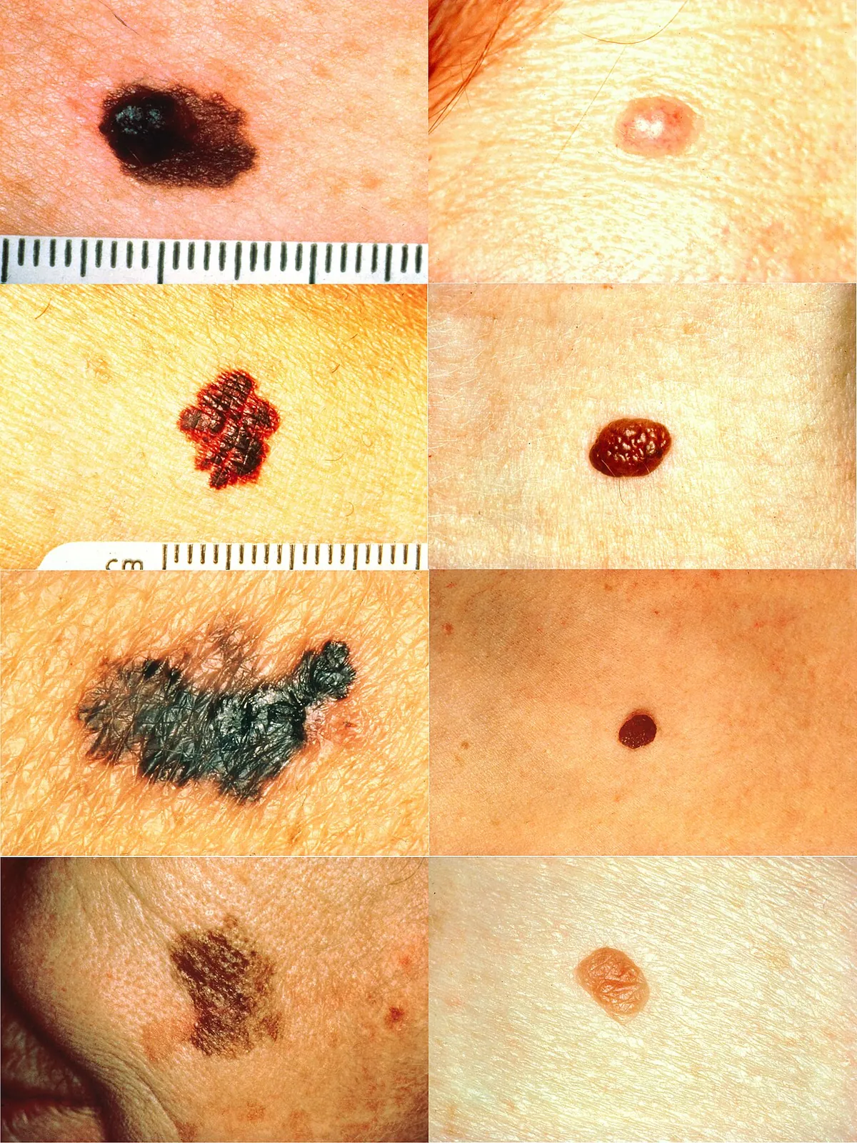
What else looks like a seborrheic keratosis?
Several common spots share the brown color or the raised, rough texture. This table draws on the AAD, DermNet and MedlinePlus; only an exam can confirm which one you have.
| Condition | What it looks like | How it differs |
|---|---|---|
| Seborrheic keratosis | Waxy, warty, "stuck-on" tan to black spot | Looks pasted on; slow to change; often many of them |
| Melanoma | Dark spot with uneven shape, border or colors | Changes in size, shape or color, can bleed; needs urgent review |
| Actinic keratosis | Dry, rough, scaly patch on sun-exposed skin | Feels like sandpaper; flatter; a precancer that needs treatment |
| Sun spot (solar lentigo) | Flat, even brown patch | Completely flat with no raised or crumbly surface |
| Common wart | Rough, skin-colored bump | Caused by a virus and can spread; often on hands |
| Mole | Even brown, round spot, flat or raised | Usually appears earlier in life; smooth surface |
| Skin tag | Soft, skin-colored flap on a stalk | Soft and smooth; hangs from the skin in folds |
For more on the flat brown spots, see our guides to melasma vs sun spots and signs of sun damage on the face. Soft hanging growths are covered in skin tags on the face.
How are seborrheic keratoses removed?
Most need no treatment at all. The AAD says a dermatologist may remove one when it looks like a skin cancer, gets caught on clothing or jewelry, becomes irritated easily, or bothers you. These are the main options, all done in a clinic:
- Shave or scrape removal: if a growth looks like skin cancer, the dermatologist will likely shave or scrape it off so a specialist can examine it under a microscope.
- Cryosurgery: liquid nitrogen is applied with a cotton swab or spray. The growth tends to fall off within days, sometimes after a blister forms and crusts over.
- Electrosurgery and curettage: the area is numbed, an electric current destroys the growth, and a scoop-shaped instrument scrapes it off. No stitches are needed.
- Other methods: DermNet also lists ablative laser and focal chemical peels.
Two side effects are worth knowing. The AAD says the treated skin may be lighter than the surrounding skin; this usually fades but is sometimes permanent. DermNet flags pigment loss as a particular concern in dark skin, so ask your dermatologist which method suits your skin tone. Most removed growths don't return, though new ones can appear elsewhere.
Insurance often treats removal for appearance alone as cosmetic, so ask about cost before the visit.
Can I remove a seborrheic keratosis at home?
No. The AAD says plainly: do not try to remove a seborrheic keratosis yourself, because of the risk of infection. Picking, cutting, freezing kits and "remover" creams also destroy the evidence a doctor needs if the spot turns out to be cancer. The FDA has told sellers that there are no over-the-counter drugs that can be legally sold for mole or skin tag removal, and the AAD warns that if a spot removed at home is a skin cancer, microscopic cancer cells can be left behind.
How can photos help you keep track?
Seborrheic keratoses change slowly, and the change that matters is often small: a new color, a bleeding edge, a spot that suddenly outgrows its neighbors. A consistent photo record is one of the easiest ways to notice. Take photos in the same light and at the same distance, and include a ruler or coin for scale when you photograph your back or chest. Our guides to lighting for skin photos and mole checker apps explain what helps.
Pori's photo diary keeps dated selfies of your face side by side. It does not assess spots, moles or growths and can't tell a keratosis from melanoma. Bring your photos to a dermatologist, who can examine the spot with a dermatoscope and biopsy it if needed.
Frequently asked questions
What does a seborrheic keratosis look like?
It is a waxy, scaly or warty growth that looks stuck onto the skin, like a dab of brown candle wax. Most are tan or brown, but they range from white to black and from a few millimeters to more than an inch across.
Can a seborrheic keratosis turn into melanoma?
Seborrheic keratoses are harmless and are not considered precancerous. The danger is that a melanoma can look like one. In one study, about 1 in 5 melanomas that resembled a seborrheic keratosis still looked like one under a dermatoscope, so any new or changing spot should be checked.
Why do I keep getting more seborrheic keratoses?
They become more common with age and tend to run in families. They are not contagious and don't spread by touch; you simply develop new ones over time. Many new ones appearing suddenly is rare and should be checked by a doctor.
How do dermatologists remove seborrheic keratoses?
Common methods are freezing with liquid nitrogen, shaving or scraping the growth off, and electrosurgery with curettage. If a growth looks like skin cancer, it is shaved or scraped so it can be examined under a microscope. Treated skin can end up lighter.
Can I remove a seborrheic keratosis at home?
No. The American Academy of Dermatology says not to remove one yourself because of the risk of infection. Home removal can also destroy the evidence a doctor needs if the spot turns out to be skin cancer, and the FDA has approved no drugs for removing these growths at home.
Are seborrheic keratoses different on dark skin?
On medium to dark skin they tend to be small, dark and often around the eyes. Many tiny dark bumps on the face and neck are often dermatosis papulosa nigra, a variant of seborrheic keratosis. Removal can leave lighter or darker marks on dark skin, so choose a dermatologist experienced with skin of color.
Sources
- American Academy of Dermatology: Seborrheic keratoses: Signs and symptoms
- American Academy of Dermatology: Seborrheic keratoses: Who gets and causes
- American Academy of Dermatology: Seborrheic keratoses: Diagnosis and treatment
- American Academy of Dermatology: Seborrheic keratoses: Tips for managing
- DermNet: Seborrhoeic keratosis
- MedlinePlus: Seborrheic keratosis
- Carrera C et al. Dermoscopic Clues for Diagnosing Melanomas That Resemble Seborrheic Keratosis. JAMA Dermatology, 2017
- American Academy of Dermatology: 5 reasons to see a dermatologist for mole, skin tag removal
- FDA: Warning letter to Amazon.com on mole and skin tag removal products (2022)
Pori is a wellness and skincare education app, not a medical device. This article is for general information and isn't medical advice. For anything that worries you, see a dermatologist.