 Pori

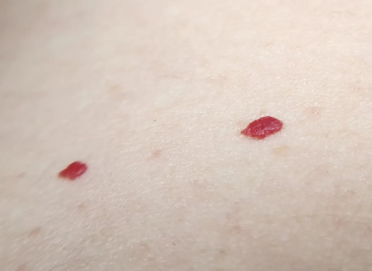
- Cherry angiomas are harmless growths of tiny blood vessels: bright red dots or smooth domes from pinhead size to about a quarter inch, usually on the trunk and arms.
- They become common after 30 and multiply with age; they tend to run in families and are not contagious or cancerous.
- Flat pinpoint dots that don't fade when pressed may be petechiae, which can be urgent with fever; a fast-growing, bleeding red lump or a dark bump needs a doctor.
- Removal is optional: electrocautery, cryotherapy, laser or shave excision all work; on darker skin ask about the risk of light or dark marks.
Cherry angiomas are small, bright red dots or smooth little domes made of tiny blood vessels. They usually appear on the chest, back and arms from about age 30, become more numerous with age, and are harmless. You only need to remove one if it bothers you, bleeds or catches on clothing, or if a doctor is unsure what it is.
Pori does not diagnose skin conditions. The selfie scans and photo diary in Pori can help you keep dated, consistent photos to show a dermatologist and to see whether a spot is changing.
Below: what cherry angiomas look like on different skin tones, why they appear, the red spots that are something else, and the ways a doctor can remove them.
What does a cherry angioma look like?
MedlinePlus describes a cherry angioma as a noncancerous (benign) skin growth made up of blood vessels. It is bright cherry red, can be flat and smooth or stick out from the skin, and ranges from pinhead size to about a quarter inch (0.64 cm) across. DermNet adds that they can be red, blue or purple, are usually multiple, and are rarely found on the hands, the feet or inside the mouth.
They don't itch or hurt. Small ones look like a drop of red ink on the skin; larger ones look like a smooth, shiny red bead. Doctors also call them Campbell de Morgan spots, senile angiomas or cherry hemangiomas, which are all the same thing.
Where on the body do they appear?
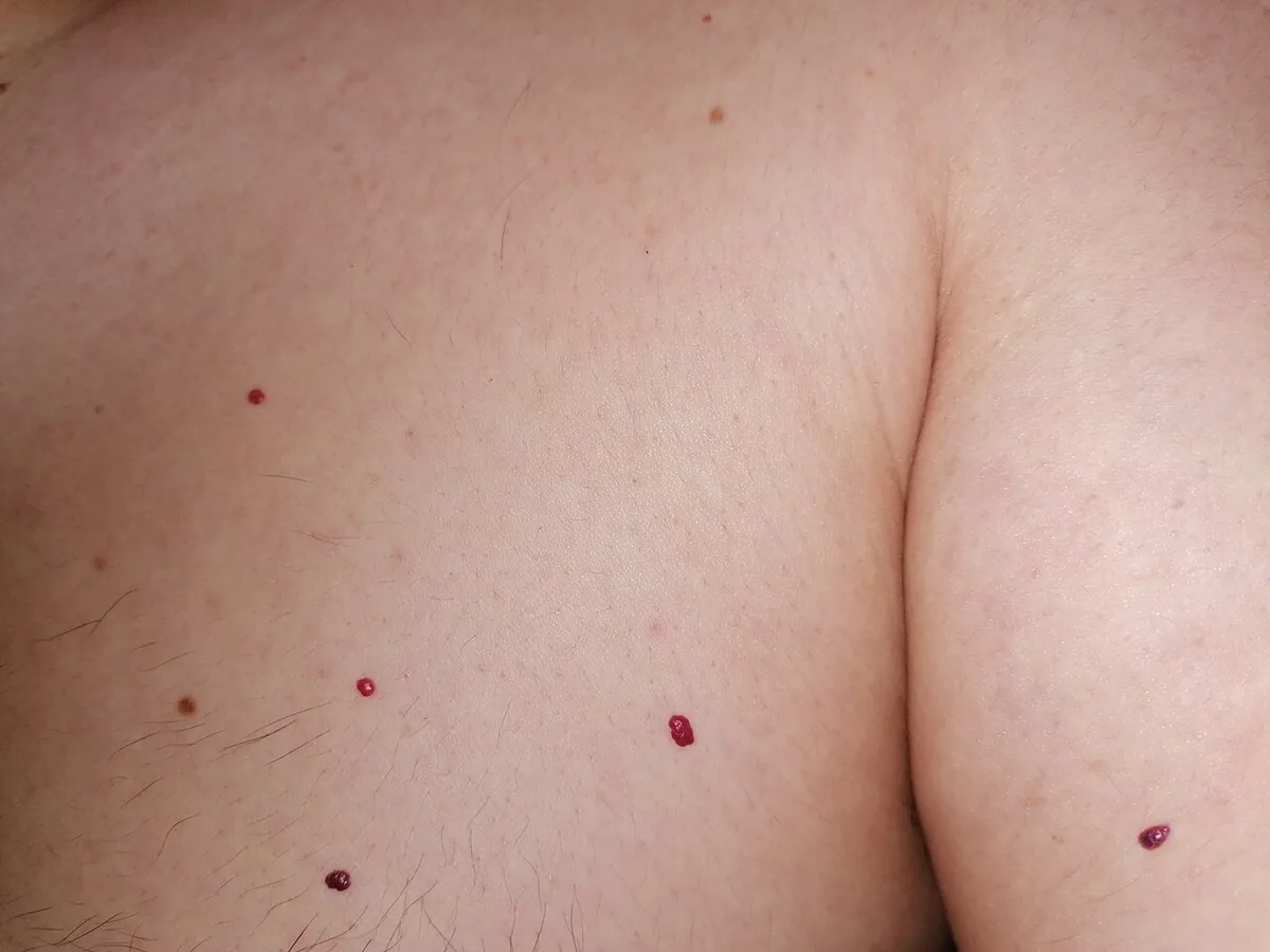
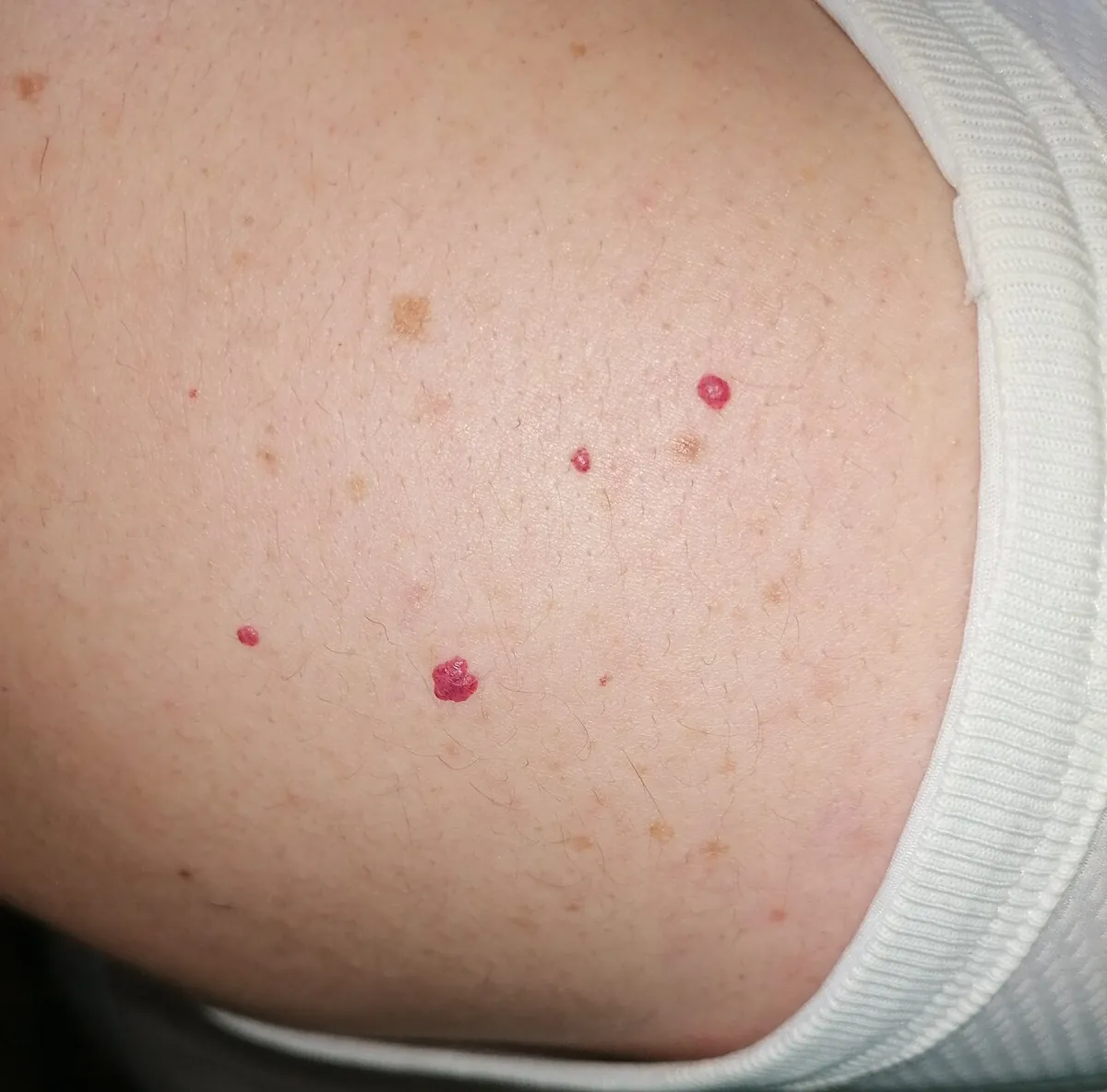
MedlinePlus says they can occur almost anywhere but usually develop on the trunk: the chest, belly and back. DermNet says they can be scattered over any part of the body, so the arms, shoulders, face and scalp are possible too; on the face and scalp they are easy to nick with a razor or comb.
How do they look on brown and black skin?
Cherry angiomas occur in people of all skin tones and sexes, but DermNet notes they are more noticeable in white skin than in skin of colour. Because a dark, raised spot on brown or black skin has a wider list of possible causes, it is worth showing a dermatologist the first time.
Why do cherry angiomas appear, and who gets them?
Age is the main factor. MedlinePlus says they are most common after age 30, and DermNet estimates that about 75% of people over 75 have them, while some teenagers get a few too. A 2020 systematic review in Dermatologic Surgery describes them as benign vascular proliferations associated with aging.
- Genes: MedlinePlus says the cause is unknown but they tend to run in families. DermNet notes that many carry changes in the GNAQ and GNA11 genes inside the spot itself.
- Pregnancy: DermNet lists pregnancy among the rare settings for a sudden crop of new angiomas.
- Sudden eruptions: DermNet notes that eruptive cherry angiomas have rarely been linked to internal cancer. If dozens appear over weeks, mention it to your doctor; a gradual increase over years is the usual pattern.
Cherry angiomas are not contagious, and they are not caused by anything you put on your skin. Because their cause is unknown and they tend to run in families, there is no proven way to prevent them. If you want to know whether new ones are appearing, a dated photo of the same area every few months is more reliable than memory.
Are cherry angiomas dangerous?
No. They are benign and do not turn into cancer. MedlinePlus says they usually do not need to be treated. The two practical problems are bleeding and doubt. Because it is made of blood vessels, a raised angioma can bleed when it is scratched or nicked while shaving. Press on it with a clean cloth until it stops. MedlinePlus lists frequent bleeding as one reason to have one removed.
The doubt is about look-alikes. DermNet notes that a cherry angioma with a clot inside can look black in colour until examined with a dermatoscope, and that doctors occasionally remove one to rule out a nodular melanoma. That is rare, but it is why a new dark or fast-growing "angioma" deserves a professional look.
When is a red spot not a cherry angioma?
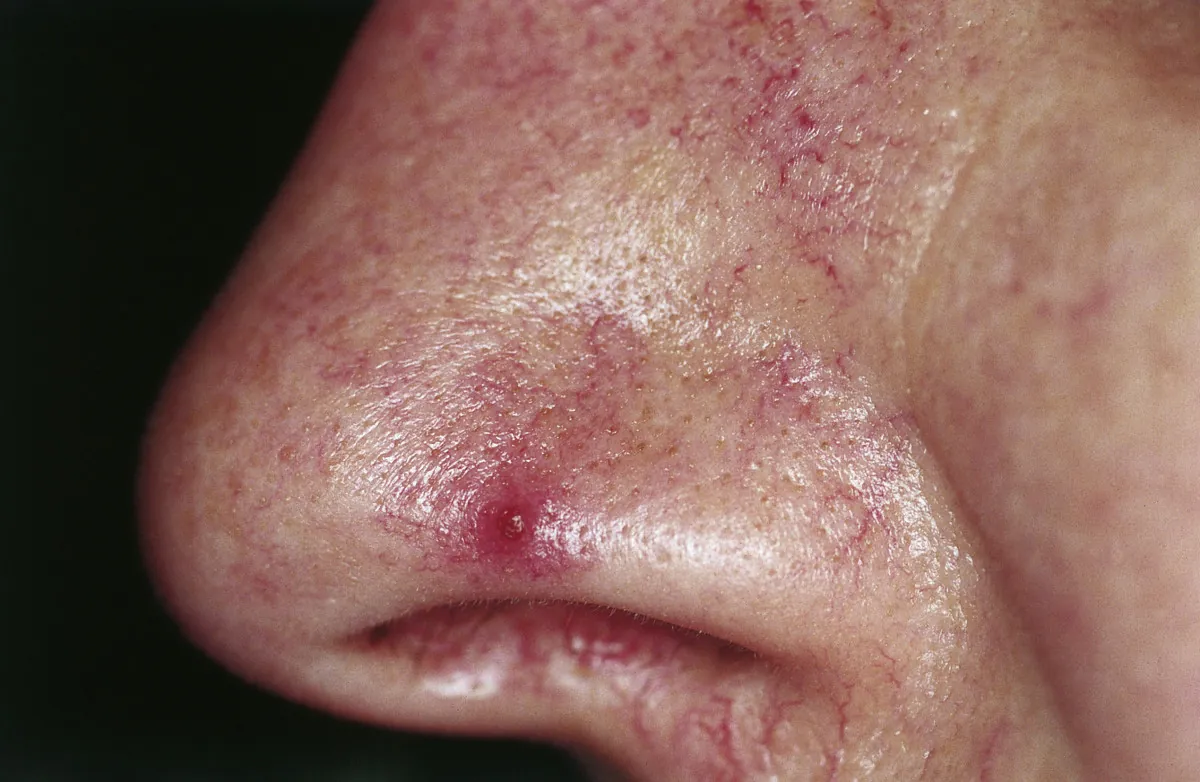
Several other red spots get mistaken for cherry angiomas. Pressure helps with two of them: petechiae stay the same color when pressed, and a spider angioma's fine legs disappear and then refill.
| Spot | How it differs from a cherry angioma |
|---|---|
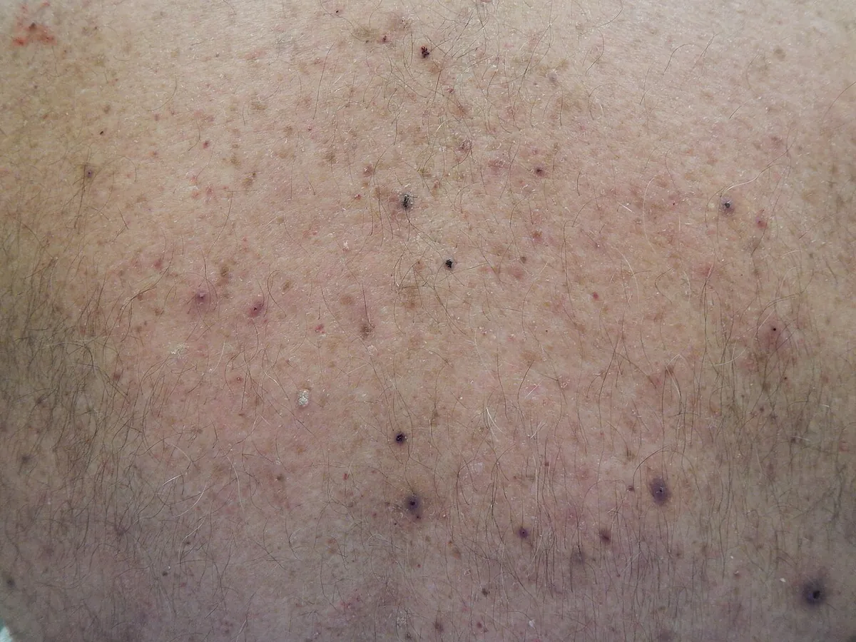
| Petechiae | Flat, pinpoint red, purple or brown dots that don't fade when pressed and often appear in clusters; can mean bleeding under the skin |
| Spider angioma | Red center with thin vessels spreading out like legs; fades when the center is pressed |
| Pyogenic granuloma | A red, fleshy lump that grows fast over a few weeks and bleeds very easily |
| Nodular or amelanotic melanoma | A firm, growing dome that can be pink, red or dark and may bleed; it keeps changing |
| Acne papule or folliculitis | Tender red bump that comes and goes within days to weeks |
Petechiae: when red dots are urgent
Cleveland Clinic describes petechiae as tiny spots of bleeding under the skin: flat purple, red or brown dots the size of a pinpoint that stay the same color when pressed. Straining, some medicines, infections and a low platelet count can cause them. Get medical care right away if pinpoint dots come with a fever, confusion, dizziness or fainting, trouble breathing, or spots that spread quickly, especially in a child.
Spider angiomas
DermNet describes a spider angioma (spider telangiectasis) as a central red papule with fine red lines spreading out from it. A single one is common in children and adults and is harmless. Several at once are most often seen in pregnancy, with birth control pills, and in people with liver disease, so many new spiders are worth a doctor's visit.
Pyogenic granuloma
DermNet describes a pyogenic granuloma as a painless red fleshy nodule, typically 5 to 10 mm, that grows rapidly over a few weeks and bleeds easily with minor bumps. It often appears after a small injury and in about 5% of pregnancies. Unlike a cherry angioma, it usually needs removal.
For red spots on the face specifically, including acne marks and rosacea, see our guide to red spots on the face. For brown and black spots that people often confuse with angiomas or moles, see seborrheic keratosis and melanoma vs a normal mole.
How are cherry angiomas removed?
Removal is optional and is often done for looks. MedlinePlus lists four methods and notes that removal usually does not cause scarring:
- Electrocautery (electrosurgery): heat from a fine electric needle seals the spot.
- Cryotherapy: freezing with liquid nitrogen.
- Laser: vascular lasers aimed at the blood vessels in the spot.
- Shave excision: the doctor shaves off a raised angioma, which also lets a lab examine it if there is any doubt.
Which removal method is best?
No single method wins. The 2020 systematic review found that laser and non-laser treatments all worked and that no therapy proved to be superior. Among lasers, pulsed dye laser caused less procedure pain than KTP laser or electrodesiccation. The Nd:YAG 1064 nm laser caused fewer pigment problems, while KTP and pulsed dye lasers risked pigment changes in people with darker skin. If you have brown or black skin, ask which method the clinic uses and how often it causes light or dark marks on skin like yours.
Treatment removes the spots you have. It does not stop new ones from forming as you get older.
Can you remove cherry angiomas at home?
We don't recommend it. Cutting, tying off or burning an angioma at home risks bleeding, infection and scarring, and it destroys the spot before anyone can check it. Online "removal pens" and acid creams are not a substitute for a diagnosis. If a spot bothers you, a short visit for electrocautery or laser is the safer route.
When should you see a doctor about a red spot?
Most cherry angiomas never need a visit. See a dermatologist if:
- A spot is new and you are not sure what it is, especially if it is dark, black or on brown or black skin
- An "angioma" grows quickly, changes shape or color, or becomes firm
- A spot bleeds often or bleeds without being injured
- Many new red spots appear over a few weeks
- Several spider-like red spots appear
- You want one removed
Seek urgent care for flat pinpoint dots that don't fade when pressed and come with fever or feeling unwell. For a skin check or removal, you can find a dermatologist near you.
Frequently asked questions
What causes cherry angiomas?
The exact cause is unknown. They are linked to aging, become common after 30, and tend to run in families. Pregnancy can bring on new ones. They are not caused by diet or skincare products.
Are cherry angiomas a sign of cancer?
No. Cherry angiomas are benign and do not turn into cancer. Very rarely, a sudden eruption of many new ones has been linked to internal disease, and a dark or fast-growing spot can be something else, so new or unusual spots are worth showing a doctor.
How can I tell a cherry angioma from petechiae?
A cherry angioma is a bright red dot or small dome that stays for years. Petechiae are flat pinpoint dots that don't fade when pressed and often appear in clusters. Petechiae with fever, confusion or fast-spreading spots need urgent care.
Why does my cherry angioma bleed so much?
It is made of blood vessels, so a scratch or razor nick can bleed a lot for its size. Press firmly with a clean cloth for several minutes. If it bleeds often, a dermatologist can remove it.
What is the best way to remove cherry angiomas?
Electrocautery, cryotherapy, laser and shave excision all work, and a 2020 review found no method clearly better. Pulsed dye laser was less painful, and Nd:YAG laser caused fewer pigment changes, which matters on darker skin.
Do cherry angiomas go away on their own?
Usually not. They tend to stay, and more may appear with age. They don't need treatment unless they bother you or bleed.
Sources
Pori is a wellness and skincare education app, not a medical device. This article is for general information and isn't medical advice. For anything that worries you, see a dermatologist.